La rete delle conoscenze nefrologiche
Attenzione! Per visualizzare al meglio il sito e usufruire di tutte le funzionalità messe a disposizione
si consiglia di aggiornare la versione in uso di Internet Explorer alla versione 8 o superiore. Grazie!
La rete delle conoscenze nefrologiche

Home > For the History of Dialysis
Pubblicato il 18 febbraio 2016
Scarica l’APP del GIN per il tuo smartphone o tablet:


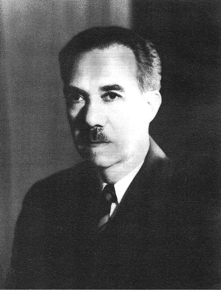
J. Stewart Cameron
Emeritus Professor of Renal Medicine. Department of Nephrology and Transplantation, Guy’s Campus, King’s College, London
Address correspondence to: J. Stewart Cameron; Elm Bank, Melmerby Cumbria CA10 1HB United Kingdom; Tel:+44 (0)1768 881804 e-mail: jstewart.cameron2@btopenworld.com
© 2013-2025 Società Italiana di Nefrologia — ISSN 1724-5990 — Editore Tesi SpA
Giornale Italiano di Nefrologia è una testata giornalistica registrata presso il Tribunale di Milano. Autorizzazione n. 396 del 10.12.2013.
La piattaforma web su cui condividere in maniera semplice, efficace ed interattiva le conoscenze nefrologiche attraverso la pubblicazione online di documenti multimediali.
NephroMEET accoglie come documenti con marchio SIN quelli approvati da: Comitati e Commissioni ufficiali SIN, Gruppi di Studio SIN, Sezioni Regionali/Interregionali SIN.
Il Consiglio Direttivo SIN si riserva inoltre la facoltà di certificare con marchio SIN altri documenti qualora lo ritenga opportuno.
Gli Autori si assumono in ogni caso la responsabilità dei contenuti pubblicati.
I contenuti pubblicati sono riservati ad un pubblico esperto nel settore medico-scientifico.
Seguici su Twitter
Developer e partner tecnologico:
TESISQUARE®
Assistenza telefonica allo 0172 476301
o via mail